Red lunula :
How the nail appears: The lunula is red. Notwithstanding the red lunula imagined here, there is additionally the nonappearance of azure lanula and lanula.
What to presume: Red lunula has been related with alopecia aerate, and collagen vascular ailment. It has additionally occurred in patients on oral prednisone for rheumatoid joint pain. Red lunulae are seen in carbon monoxide harming, psoriasis, chronic urticaria, cirrhosis, COPD and cardiovascular breakdown.
Nonappearance of lunula was the most widely recognized nail issue in a gathering of hemodialysis patients (over 30%) and has likewise been accounted for in renal transplant beneficiaries (over 17%). Azure lunula is also noticed in patients with Wilson illness. It has also been seen in SNS nail dipping powder kit, agrarian and in patients taking pharmaceuticals like azidothymidine and 5-fluorouracil.
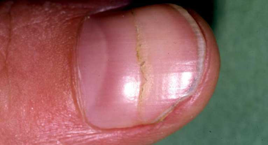
Chip haemorrhages:
How the nail appears: Extravasations of blood from the longitudinally situated vessels of the nail bed. These haemorrhages don’t whiten. They shape because of the nail plate-dermis auxiliary relationship and have a tendency to be seen in older patients not using sns powder nails.
What to presume: While injury is the most widely recognized reason, occur with fungal infection and psoriasis.
Bacterial endocarditis a most well-known fundamental illness related with chip haemorrhages. These haemorrhages are more typical in subacute, as opposed to intense diseases. Despite the fact that chip haemorrhages in subacute bacterial endocarditis have been depicted as proximally situated, there are no adequate information to affirm this—for the most part since fragment sores move distally as the nail develops.
Chip haemorrhages may likewise be related with Darier’s disease, chronic glomerulonephritis, scurvy, trichinosis, cirrhosis, vasculitis and mitral stenosis. Nonetheless, due to the assorted and basic reasons for fragment haemorrhages, they can’t be utilized as a segregated indication of some diseases, with the exception of when they are accompanied by things like heart murmur, Janeway’s lesions, Osler’s nodes, Roth’s spots, fever, since any of the above would incredibly build their criticalness.
Terry’s nails:
How the nails appear: Most of the nail plate appears white, with a limited pink distal band. All nails have a tendency to be consistently affected, with an appearance of ground glass. Terry’s nails have been found in over 70% of patients with cirrhosis of the liver.
What to presume: One examination discovered Terry’s nails in 30% of 627 patients, with analysts connecting the symptoms with diabetes mellitus, chronic CHF and cirrhosis. Rarely, Terry’s nails have been accounted for in hemodialysis patients and renal transplant beneficiaries. Terry’s nails have additionally been seen in HIV patients.
Koilonychia:
How the nail appears: Concave thin nails with averted edges formed like a spoon and fit for holding a drop of water. It is more typical in fingernails, however is sometimes found in toenails with sns dip colors.
What to presume: This nail sign may come about because of injury, consistent introduction of hands to oil based solvents, or nail-patella disorder. Koilonychia is most generally connected with iron deficiency and incidentally happens in patients with hemochromatosis. Other popular reasons for koilonychia are hypothyroidism and coronary disease. Moreover, koilonychia is often a rare variation in much younger children and vanishes as they grow older.